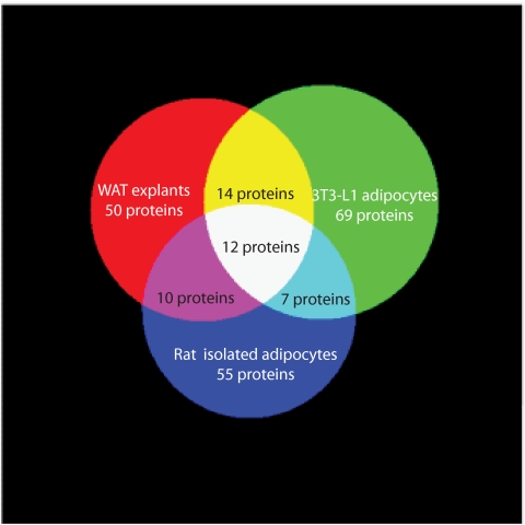
FIG. 4.
Venn diagram showing overlap of protein secretion from WAT explants, 3T3–L1 adipocytes, and isolated rat adipocytes (from ref 27). The 12 proteins detected in all three samples were adiponectin, adipsin, angiotensinogen (SerpinA8), cathepsin B, cathepsin D, collagen α-1(IV), collagen α-2(IV), complement C1s, haptoglobin, laminin subunit β-2, osteonectin, and thrombospondin-1. The diagram was composed using 3-Venn applet (48). (A high-quality color representation of this figure is available in the online issue.)

