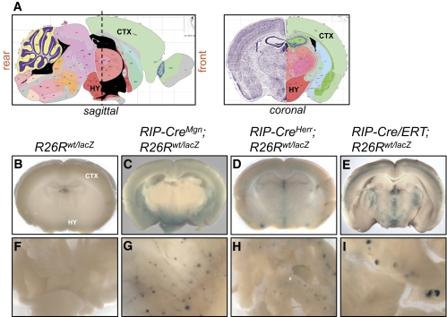
FIG. 1.
RIP-Cre transgenic lines display Cre-mediated recombination in multiple regions of the brain. Adult brains were sliced into four or five coronal sections and subjected to whole mount X-gal staining. Images of individual brain slices from each sectioning plane are available in supplementary Figs. 1–4. A: Sagittal and coronal views of mouse brain (adapted from Allen Mouse Brain Atlas, http://www.brain-map.org/) (29). A dashed vertical line marks coronal sectioning plane spanning the hypothalamic region of the brain. B–E: Images of coronal brain slices located on the left side of the sectioning plane in the sagittal view in A. B: R26Rwt/lacZ littermate control mice (n = 17) lacked X-gal staining in the brain. The cortex (CTX) and hypothalamus (HY) are labeled and correspond to regions marked on the coronal view in A. C: RIP-CreMgn;R26Rwt/lacZ mice (n = 8) showed X-gal staining throughout the brain with high signal intensity in the mid-brain and ventral regions. D: RIP-CreHerr;R26Rwt/lacZ mice (n = 14) showed weaker, punctate X-gal staining throughout the brain without obvious regionalization. E: RIP-Cre/ERT;R26Rwt/lacZ mice (n = 4) injected intraperitoneally with three 2-mg doses of tamoxifen over a 5-day period displayed strong, punctate X-gal staining throughout the brain with expression pattern more restricted than in RIP-CreMgn; R26Rwt/lacZ mice. Brains from littermate controls injected with corn oil vehicle were negative for X-gal staining (data not shown). F–I: Whole-mount X-gal staining of pancreas from R26Rwt/lacZ in F, RIP-CreMgn; R26Rwt/lacZ in H, RIP-CreHerr;R26Rwt/lacZ in G, and RIP-Cre/ERT;R26Rwt/lacZ mice in I. (A high-quality digital representation of this figure is available in the online issue.)

