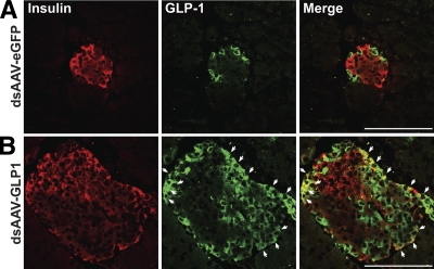
FIG. 3.
dsAAV-expressed GLP-1 is localized to insulin-producing β-cells. Expression of GLP-1 in db/db mice treated with dsAAV–eGFP (A) and dsAAV–GLP1 (B) was examined by immunofluorescence at 14 weeks of age, or 10 weeks post-treatment. Islets from mice treated with dsAAV–GLP1 contained examples of β-cells (indicated by white arrows) that were positive for both GLP-1 (green) and insulin (red). Islets from mice treated with dsAAV–eGFP contained no examples of GLP-1 and insulin double-positive cells, indicating that the green staining observed in these samples is glucagon from pancreatic α-cells. Data are representative of at least two sections per mouse, n = 10 mice per group. Scale bar = 100 μm in all images. (A high-quality digital representation of this figure is available in the online issue.)

