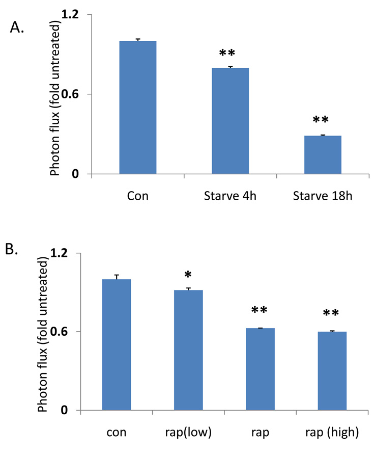
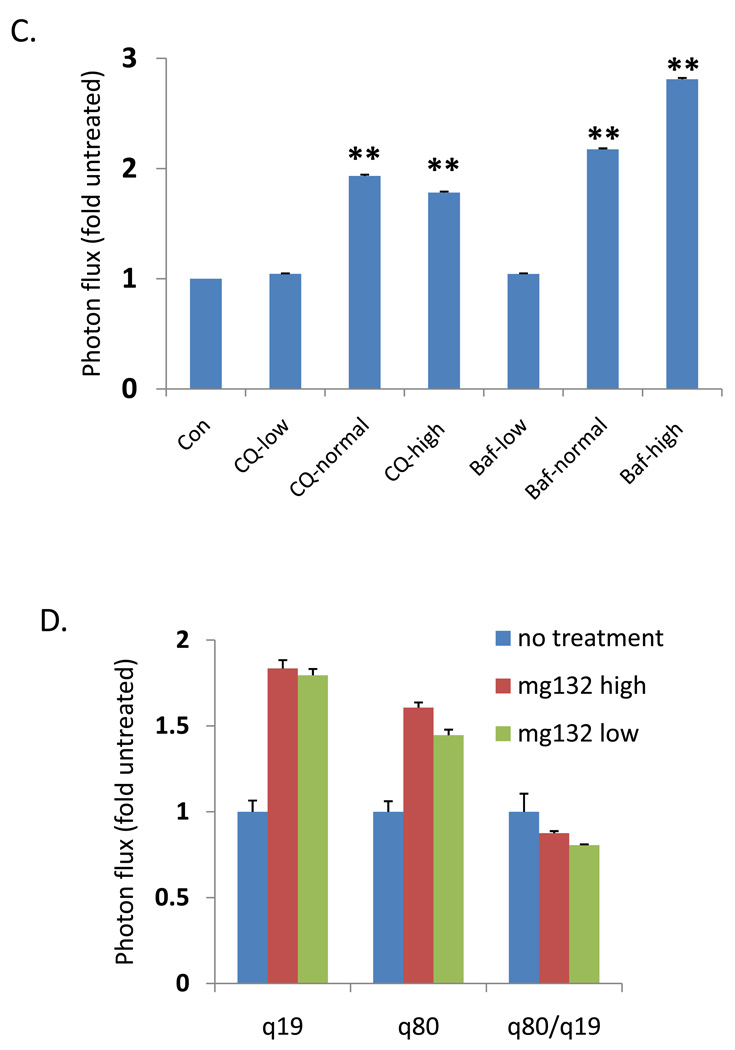
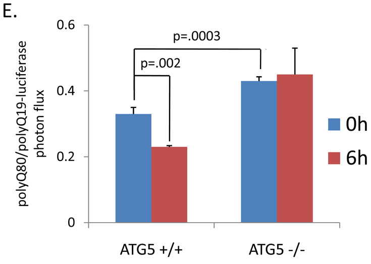
Figure 3.
The ratio of polyQ80/polyQ19-luciferase activity in differentiated C2C12 myotubes transfected following (A) starvation for 4 or 18 hours (B) increasing concentrations of rapamycin for 18 hours or (C) increasing concentrations of autophagy inhibitors chloroquine (CQ) or bafilomycinA (Baf). * denotes a p value of <.01. ** denotes a p value of <.001. (D) U20S cells treated with proteasome inhibitor MG132 have a similar increase in both Q19 and Q80-luciferase activity after 4 hours of treatment. E) ATG5+/+ or ATG5−/− mouse embryonic fibroblasts (MEFs) were transfected with polyQ80 or polyQ19-luciferase and subjected to nutrient deprivation for 0 or 6 hours. The raw ratio of polyQ80/polyQ19-luciferase activity is presented. Note that the amount ratio of polyQ80/polyQ19-luciferase is elevated in ATG5−/− cells at time 0. In addition, whereas the ratio decreases in ATG5+/+ MEFs following nutrient deprivation, this does not occur in ATG5−/− MEFs suggesting impaired autophagic degradation.


