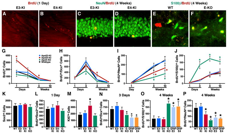
Figure 2. Effects of ApoE Deficiency and ApoE Isoforms on Hippocampal Neurogenesis and Astrogenesis.
(A, B) Representative confocal images of the BrdU-positive cells in the SGZ of female apoE3-KI (A) and apoE4-KI (B) mice at 6–7 months of age were collected 1 day after BrdU injection.
(C, D) Representative confocal images of the BrdU and NeuN double positive cells in the SGZ of female apoE3-KI (C) and apoE4-KI (D) mice at 6–7 months of age were collected 4 weeks after BrdU injection.
(E, F) Representative confocal images of the BrdU and S100β double positive cells in the SGZ of female wildtype (E) and apoE-KO (F) mice at 6–7 months of age were collected 4 weeks after BrdU injection.
(G–J) Numbers of newborn cells (BrdU+) (G), immature neurons (BrdU+/Dcx+) (H), mature neurons (BrdU+/NeuN+) (I), and astrocytes (BrdU+/S100β+) (J) in the SGZ of female mice of various apoE genotypes at 6–7 months of age were determined 1 and 3 days and 4 and 10 weeks after BrdU injection. Values are mean ± SD (n = 4–6 mice per genotype). * p < 0.05 vs. other groups (t test).M
(K) Total numbers of Sox2-positive cells in the SGZ of female wildtype, apoE3-KI, apoE4-KI, and apoE-KO mice at 6–7 months of age. Values are mean ± SD (n = 4 mice per genotype).M
(L) Numbers of BrdU and Sox2 double-positive cells in the SGZ of female wildtype, apoE3-KI, apoE4-KI, and apoE-KO mice at 6–7 months of age were determined 1 day after BrdU injection. Values are mean ± SD (n = 4 mice per genotype).
(M) Total numbers of Ki67-positive cells in the SGZ of female wildtype, apoE3-KI, apoE4-KI, and apoE-KO mice at 6–7 months of age. Values are mean ± SD (n = 4 mice per genotype).
(N–P) Numbers of immature neurons (BrdU+/Dcx+) (N), astrocytes (BrdU+/S100β+) (O), and mature neurons (BrdU+/NeuN+) (P) in the SGZ of female mice of various apoE genotypes at 6–7 months of age were determined at 3 days and 4 weeks after BrdU injection. Values are mean ± SD (n = 4–6 mice per genotype). * p < 0.05 vs. wildtype and apoE3-KI mice (t test).

