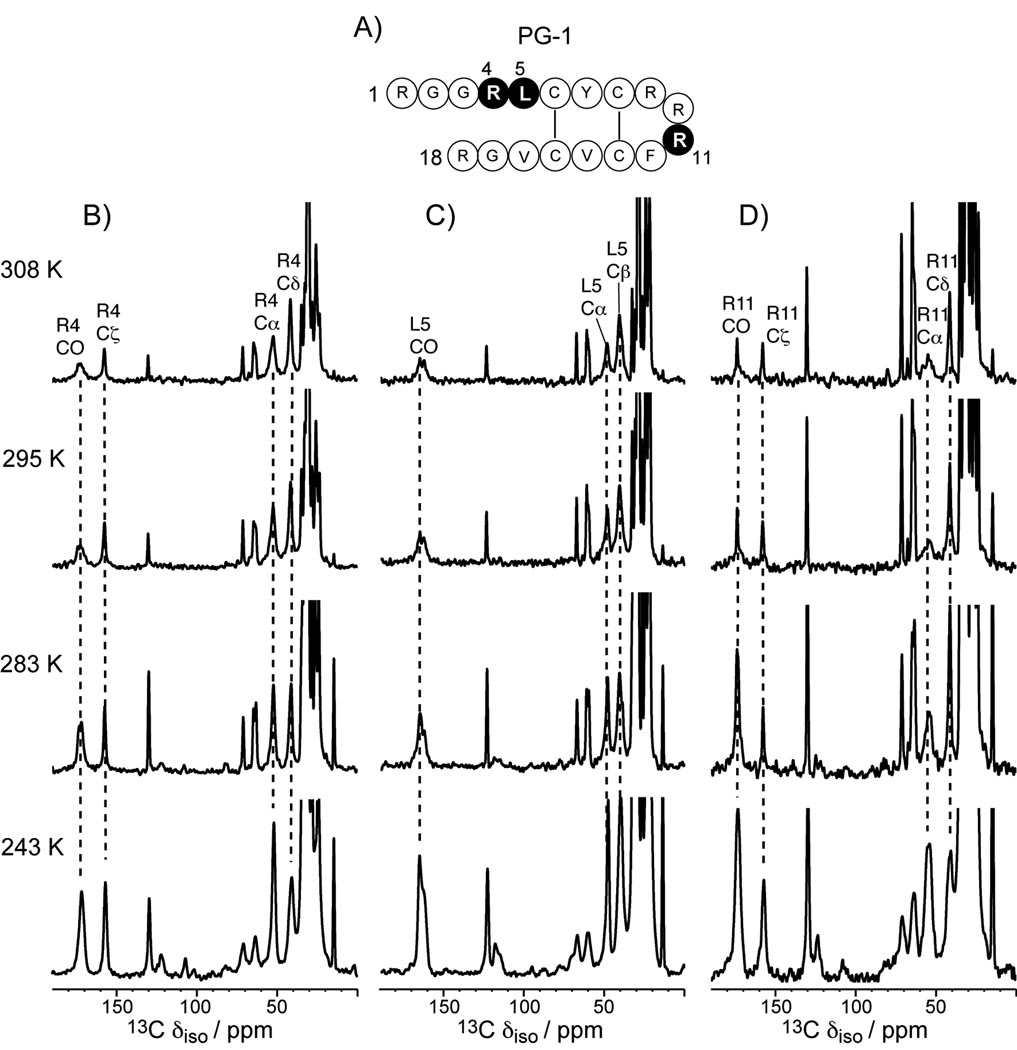
Figure 1.
13C CP-MAS spectra in PG-1 bound to the POPE/POPG membrane (P/L = 1:12.5) from 243 K to 308 K. A) Amino acid sequence of PG-1. Labeled residues are shaded. B) 13C CP-MAS spectra of Arg4, C) 13C CP-MAS spectra of Leu5, D) 13C CP-MAS spectra of Arg11. Peptide peaks are assigned and annotated.

