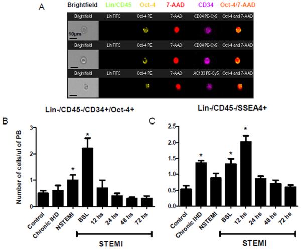
Figure 3.
Mobilizations of Oct-4 positive pluripotent VSELs in ischemic heart disease patients and controls. Panel A. Representative Image Stream pictures of circulating Oct-4 positive VSELs lacking the expression of hematopoietic lineages (Lin) and CD45 markers (Green) and positive for Oct-4 (yellow) and CD34 or CD133 (magenta). Nuclei are stained with 7-AAD (red). The combined image in the far right demonstrates the co-localization of Oct-4 in the nucleus. Panel B. Bar graphs showing the absolute numbers of circulating Lin-/CD45−/CD34+/Oct-4+ cells in the peripheral blood of ischemic heart disease patients and controls; showing a peak mobilization early in STEMI patients. Panel C. Bar graphs showing the absolute numbers of circulating Lin-/CD45−/SSEA-4+ cells in the peripheral blood of ischemic heart disease patients and controls; showing a peak mobilization early in STEMI patients. (* P < 0.05 as compared to controls). NSTEMI, non-ST-elevation myocardial infarction; PB, peripheral blood; STEMI, ST-elevation myocardial infarction.

