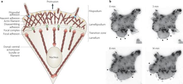
Figure 1. Structural elements of a migrating cell.
a | Adhesion is closely coupled with the protrusions of the leading edge of the cell (filopodia and lamellipodia). Adhesions (nascent adhesions) initially form in the lamellipodium (although adhesions may also be associated with filopodia) and the rate of nascent adhesion assembly correlates with the rate of protrusion. Nascent adhesions either disassemble or elongate at the convergence of the lamellipodium and lamellum (the transition zone). Adhesion maturation to focal complexes and focal adhesions is accompanied by the bundling and cross-bridging of actin filaments, and actomyosin-induced contractility stabilizes adhesion formation and increases adhesion size. b | TIRF micrographs of a Chinese hamster ovary (CHO) cell expressing paxillin–mEGFP (monomeric enhanced green fluorescent protein) on glass coated with fibronectin (5 μg ml−1). Images were acquired every 5 seconds, and representative images from 0, 3, 8 and 14 minutes are shown (see REF. 49). Closed arrow heads denote nascent adhesions assembling and turning over in protrusions. Open arrow heads indicate maturing adhesions that begin to elongate centripetally (that is, towards the cell centre) when protrusion pauses or halts. For a movie of this experiment see supplementary information S1 (movie).

