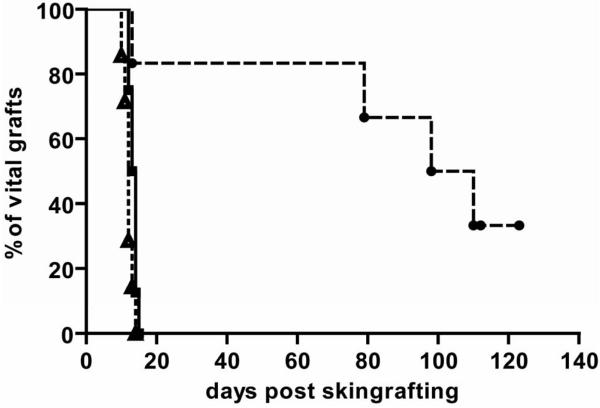
Figure 2. Donor skin graft survival is significantly prolonged in the absence of NKT cells.
Groups of mice received 1 Gy TBI with costimulation blockade and were grafted with donor and third party skin; donor skin graft survival for WT→WT (■ solid line; n= 8); donor skin graft survival for KO→KO (● slashed line; n= 6). Absence of NKT cells was associated with significantly prolonged survival of donor skin (p= 0.004 by log-rank test). Percent graft survival is shown for third-party (△ dotted line; pooled data from both groups; n= 14) and donor skin as estimated by the Kaplan-Meier product limit method.

