
Figure 2. Analysis of phosphorylation of RNF2.
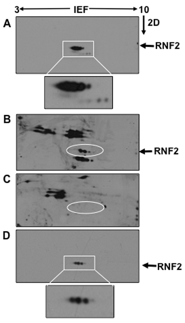
RNF2-6His was expressed in Sf9 insect cells and purified by Ni-NTA agarose beads. A. RNF2-6His was eluted from the beads and separated by isoelectric focusing (IEF) using pH3–10 IEF strips and separated by SDS-PAGE in the second dimension (2D). Proteins thus separated were transferred onto PVDF membranes followed by western blotting using anti-RNF2 antibody. RNF2-6His was resolved into multiple protein spots all of which migrated as ~39 kDa proteins. Boxed region was enlarged to show details of RNF2 spots, which was inserted below the Panel. B. An identical blot of the Panel A was developed with anti-phosphoserine antibody. RNF2-6His protein spots were shown in the circle. C. RNF2-6His eluted from the beads was dephosphorylated with protein phosphatase and then separated by 2D followed by western blotting using anti-phosphoserine antibody. The position of RNF2-6His in this blot was identified with a circle. D. The dephosphorylated RNF2 separated by 2D electrophoresis was analyzed by western blotting using anti-RNF2. Boxed region was enlarged to show details of RNF2 spots, which was inserted below the Panel.