
Figure 1.
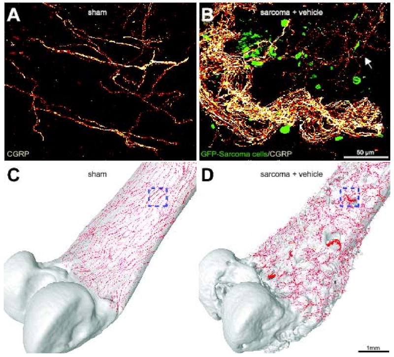
Sensory nerve fibers sprout and form neuroma-like structures as tumor cells invade the periosteum of the bone. Confocal images of non-decalcified whole mount preparations of the femoral periosteum from sham + vehicle (A) or sarcoma + vehicle mice (B) showing CGRP+ nerve fibers and GFP+ sarcoma cancer cells. When GFP+ tumor cells invade the periosteum, they induce ectopic sprouting of CGRP+ sensory fibers (B, arrow) and the formation of neuroma-like structures. Confocal images of periosteum (approximately 70 μm in thickness) were acquired from whole mount preparations and projected into a Z-stack from 280 optical sections at 0.25 μm intervals with a 40× objective. Four Z-stack images from sham or sarcoma + vehicle mice were tiled and overlayed (to scale) on a three-dimensional micro-CT rendering of a sham femur (C) or sarcoma + vehicle femur (D), respectively, using Amira software. The boxed areas in (C) and (D) correspond to the confocal images in (A) and (B), respectively. Note that the tumor-injected femur (D) has severe cortical bone deterioration and a pathological reorganization of CGRP nerve fibers (in red) compared to the sham bone (C).