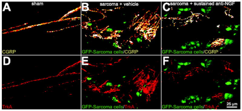
Figure 2.

Preventive sequestration of NGF reduces CGRP+ and TrkA+ nerve fiber sprouting and the formation of neuroma-like structures in the periosteum of tumor-injected mice. Representative confocal images of femoral sections from sham + vehicle (A, D), sarcoma + vehicle (B, E), and sarcoma + early/sustained anti-NGF (C, F) mice. Decalcified bone sections were double-immunostained with an antibody against CGRP (orange in A, B, C) and an antibody against TrkA (cognate receptor for NGF, red in D, E, F). Confocal images were acquired in the proximal metaphyseal periosteum (∼2 mm below the growth plate) using a sequential acquisition mode to reduce bleed-through. Note that at day 20 post-injection there is remarkable sprouting by CGRP+ nerve fibers in sarcoma + vehicle mice (B) and that nearly all the sprouted CGRP+ nerve fibers also express TrkA (E). Nerve fibers that undergo sprouting have a remarkable pathological and disorganized morphology as compared to nerve fibers innervating sham bones. Preventive sequestration of NGF (10 mg/kg; i.p., given at days 6, 12, and 18 post cell injection) significantly reduces the pathological tumor-induced reorganization of sensory CGRP+ (C) and TrkA+ (F) nerve fibers. Confocal images were acquired from bone sections (20 μm in thickness) and were projected from 120 optical sections at 0.25μm intervals with a 40× objective.
