
Figure 4.
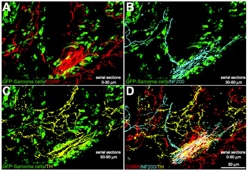
Sensory and sympathetic nerve fibers are observed in the same neuroma-like structures in tumor-bearing mice. Confocal images of serial sections of bone (each 30 μm apart) immunostained with CGRP (serial section 1, red, A), NF200 (serial section 2, blue, B), and TH (serial section 3, yellow, C) from a tumor-bearing mouse. At 20 days post-injection, GFP+ cancer cells (green, A, B, C) had induced dramatic sprouting and formation of neuroma-like structures by CGRP+, NF200+, and TH+ nerve fibers in the periosteum/cortical bone interface. Note that when the confocal images of nerve fibers are merged (D), all three fiber types appear to be present in the same neuroma-like structure, which appears white in (D). The GFP background in A to C was acquired from the middle section (serial section 2). The merged image is a Z-stack of confocal images projected from 360 optical sections at 0.25μm intervals with a 40× objective.