Fig. 1.
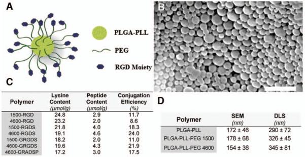
Design, synthesis and characterization of synthetic platelets. (A) Schematic of synthetic platelet comprised of PLGA-PLL core with PEG arms terminated with the RGD moiety. (B) Scanning electron microscope (SEM) micrograph of synthetic platelets. Scale bar, 1 μm. (C) Lysine and peptide concentrations of synthetic platelets as determined by amino acid analysis. Conjugation efficiency was defined as the peptide to lysine ratio multiplied by 100. (D) Diameter of PLGA-PLL core and PLGA-PLL-PEG nanospheres as determined by SEM microscopy and dynamic light scattering (DLS). SEM diameter based on n = 80. Data are expressed as mean ± SD.

