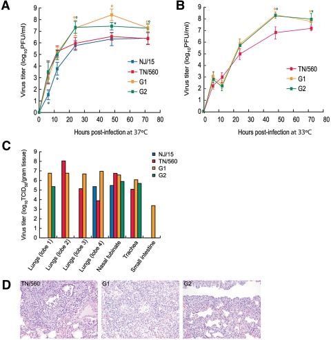
FIG 2 .
Replication of NJ/15, TN/560, and two recovered H1N1 genotypes, G1 and G2, in NHBE cells and ferrets. (A and B) NHBE cultures were infected via the apical side with each virus at an MOI of 0.1 at either 37°C (A) or 33°C (B). The progeny viruses released from the apical surfaces of infected cultures were collected at the indicated time points and titrated in MDCK cells by performing a plaque assay. Representative results expressed as log10 numbers of PFU/ml from three independent experiments are shown. *, P < 0.05; °, P <0.01 compared with the value for TN/560 virus (one-way ANOVA). (C) Four ferrets were intranasally inoculated with 106 PFU of each virus, and 3 days later, tissues were collected and virus titers were determined. Virus titers are expressed as log10 TCID50/gram tissue for one ferret. (D) The images shown are hematoxylin-and-eosin-stained sections of lung tissue from donor ferrets inoculated with TN/560, G1, or G2 (H1N1) influenza viruses obtained on day 3 postinoculation. The TN/560 lung infection was primary restricted to the bronchioles and consisted of epithelial necrosis and regeneration associated with intraluminal sloughed epithelial cells and mononuclear inflammatory cells. The G1 infection involved the bronchioles and alveoli and consisted of bronchiolar epithelial necrosis and regeneration associated with alveolar pneumocytes hyperplasia and mononuclear inflammatory cell infiltrates. The pathologic changes in the G2-infected lungs were restricted to the alveolar interstitium only. The alveolar spaces were free of inflammatory cells, whereas the alveolar interstitial walls were hypercellular with mononuclear cells. Magnification, ×20.

