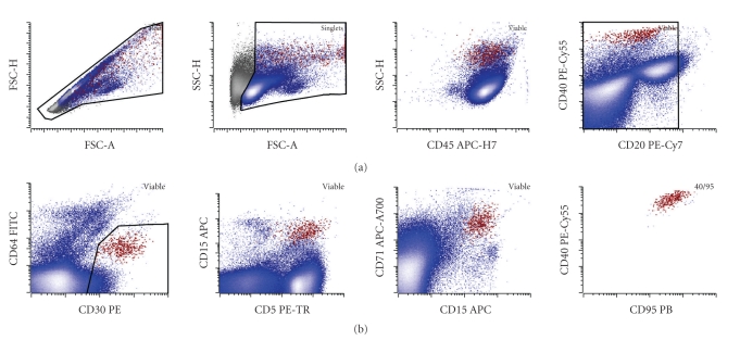
Figure 3.
Identification of a small HRS population in a clinical sample: malignant HRS cells shown in red have increased forward scatter area (FSC-A) and height (FSC-H) (note a subpopulation with HRS cells with disproportionately increased FSC-A likely corresponding to a rosetted population) and increased side scatter height (SSC-H) compared to the rest of the node cellularity (blue). The population showed bimodal, but mostly bright, expression of CD45, low levels of a B cell-marker CD20, no significant expression of a monocyte-marker CD64 (mild increase in apparent CD64 expression is due to autofluorescence of the HRS cells, which was previously verified by isotype control, data not shown), bright CD30, mostly bright CD5 (with a noticeable nonrosetted CD5 low to negative population), bright CD15 at a level slightly lower than granulocytes, and bright CD71 (transferrin receptor) consistent with high metabolic activity. Finally, the population shows a tight cluster in multiple projections including CD95 (bright) versus CD40. In this case, the HRS population represented 0.09% of the total white cells in the lymph node. The case was morphologically confirmed as CHL-nodular sclerosis type.

