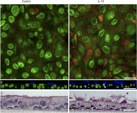
Figure 1.
Induction of mucous metaplasia by IL-13. (A) Detection of mucus with fluorescent periodic acid Schiff (orange). Focal plane, ∼5 μm below apical membrane; nuclei are green. (B) Representative staining of goblet cells with monoclonal antibody B6E8 (blue). Nuclei are stained green with Yo-Pro-1. (C) Light microscopy with mucicarmine staining. Distended cell bodies of several goblet cells are marked by asterisks. Several more lie predominantly outside the plane of the section, but all can be identified by the mucus they release. Each pair of images was from cell sheets derived from the same trachea. Scale bars = 25 μm (A), 50 μm (B), and 20 μm (C).

