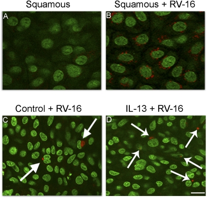
Figure 4.
Detection of rhinovirus infection of airway epithelial cells by rhinovirus (RV)-16 immunohistochemistry (nuclei, green, using Yo-Pro-1; RV-16, red). (A) Squamous-cell cultures not receiving virus. (B) Squamous cultures exposed to RV-16. (C) Mucociliary cultures exposed to virus demonstrate occasional perinuclear staining of infectious particles (arrows). (D) IL-13–treated cells exposed to virus. Cell sheets are from the same tissue. Scale bar = 10 μm.

