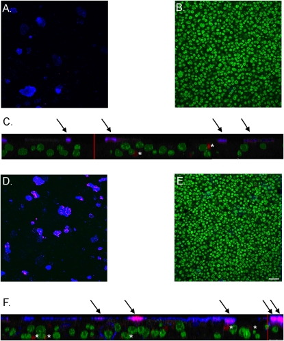
Figure 5.
Rhinovirus preferentially infects goblet cells, an effect enhanced by goblet-cell metaplasia induced by IL-13. Nuclei, green; mucus, blue; virus, red. (A) Apical plane of control cell sheets shows colocalization of virus to goblet cells. (B) Center plane of control cell sheets shows no mucus staining. (C) Z-stack of control cell sheets. (D) Apical plane of IL-13–treated cell sheet shows colocalization of virus to goblet cells. (E) Center plane of IL-13–treated cell sheet shows no mucus staining. (F) Z-stack of IL-13–treated cell sheets; scale bar = 25 μm. Arrows indicate colocalization of mucus and virus. Where this occurs in IL-13–treated cells, levels of both mucus and virus are higher than in control cells. Asterisks indicate virus unassociated with mucus. Cells sheets were grown from the same trachea. Scale bar in E = 25 μm. Same scale for A, B, D, and E. Red vertical bars in z-stacks represent 25 μm.

