Figure 7.
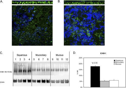
Quantification of rhinovirus receptor ICAM-1 in airway epithelial cells. (A) Immunofluorescence of ICAM-1 in control cell sheets shows the receptor is primarily localized to the apical surface. (B) Immunofluorescence of ICAM-1 in IL-13–treated cell sheets shows similar apical localization of ICAM-1, with no apparent association with goblet-cell staining. Top: Z-projection of an x–y stack. Bottom: x–y plane at level of the apical membrane. B6E8, blue; ICAM-1, green, at apical membrane; nuclei, green, using Yo-Pro-1. Scale bar = 15 μm. (C) Squamous-cell sheets have higher levels of ICAM-1 protein levels, compared with control cell sheets when normalized to β-actin levels. Mucus (IL-13–treated) and mucociliary cell sheets have similar levels of ICAM-1 protein when normalized to β-actin levels. (D) Pooled data from three experiments. AU, arbitrary units as a measure of pixel density.

