Figure 7.
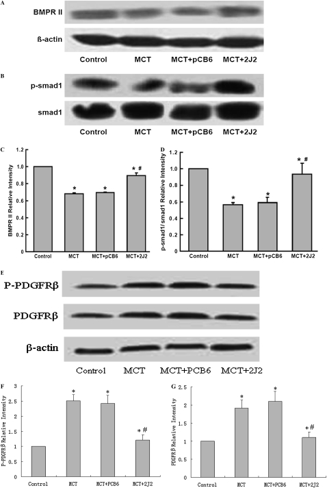
Bone morphogenetic protein (BMP) and platelet-derived growth factor receptor β (PDGFRβ) signaling in MCT-treated rat lungs assessed by Western blotting. A significant decrease occurred in BMPRII (A) and p-Smad1 (B) expression in rats treated with MCT, either alone or together with pCB6 empty vector. However, in MCT-treated rats receiving pCB6-2J2, concentrations of BMPRII and p-Smad1 protein were increased. Results are representative of three independent experiments. Blots were scanned, and relative BMPRII (C) and p-Smad1 (D) were normalized to β-actin and Smad1, respectively. (E) Concentrations of PDGFRβ and phosphorylated PDGFRβ were upregulated in MCT-treated animals, and treatment with CYP2J2 attenuated the upregulation. (F and G) Relative PDGFRβ and phosphorylated PDGFRβ, respectively, were normalized to β-actin. Data shown are mean ± SEM of 6– 8 rats/group. *P < 0.05 versus control. #P < 0.05 versus MCT + pCB6.

