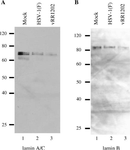
Fig. 10.
Lamin disruption in infected HEp-2 cells is associated with loss of lamin protein. Shown are digital images of western blots of protein from HEp-2 cells infected for 24 h probed for lamin A/C (A) and lamin B (B). Coomassie staining was used to equilibrate loading (not shown). These experiments were done three times. Representative images are shown. The infecting virus is indicated above each lane.

