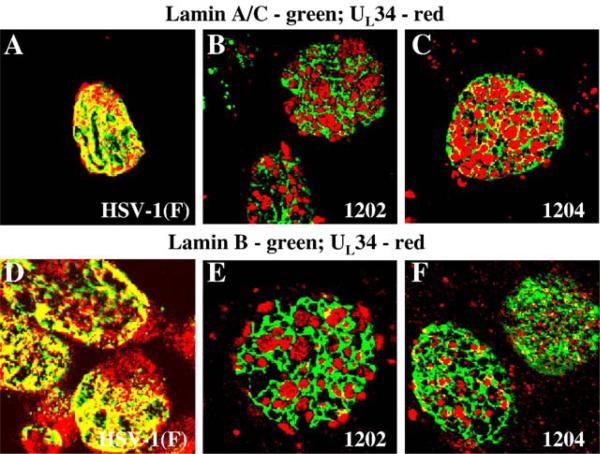
Fig. 4.
Association of UL34 with lamin perforations in cells infected with US3 mutant viruses. Shown are digital confocal images showing the localization of UL34 and lamin A/C or lamin B in infected Vero cells. Green: lamins, red: UL34. Vero cells were infected with HSV-1(F) (A and D) vRR1202 (B and E) or vRR1204 (C and F) for 24 h at an MOI of 5. Cells were stained for UL34 (A–F) and for either lamin A/C (A–C) or lamin B (D–F).

