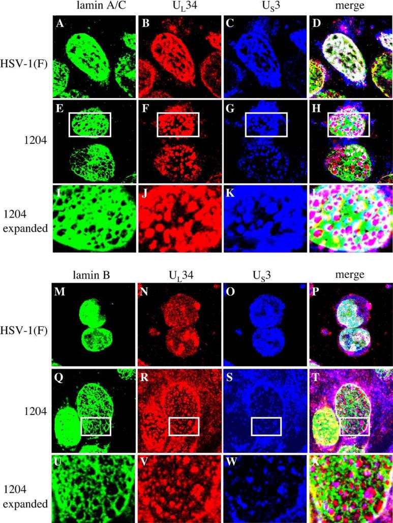
Fig. 5.
Association of US3 with lamin perforations in cells infected with kinase-dead US3 mutant virus. Shown are digital confocal images of UL34, US3 and lamin A/C or lamin B localizations in HSV-1(F) or vRR1204 infected Vero cells. Green: lamins, red: UL34, blue: US3. For all panels, Vero cells were infected with the HSV-1(F) or vRR1204 for 24 h at an MOI of 5. Infecting viruses are indicated to the left of the figure. Panels I–L and U–X each show an enlarged view of the area enclosed in the white box in the panel immediately above.

