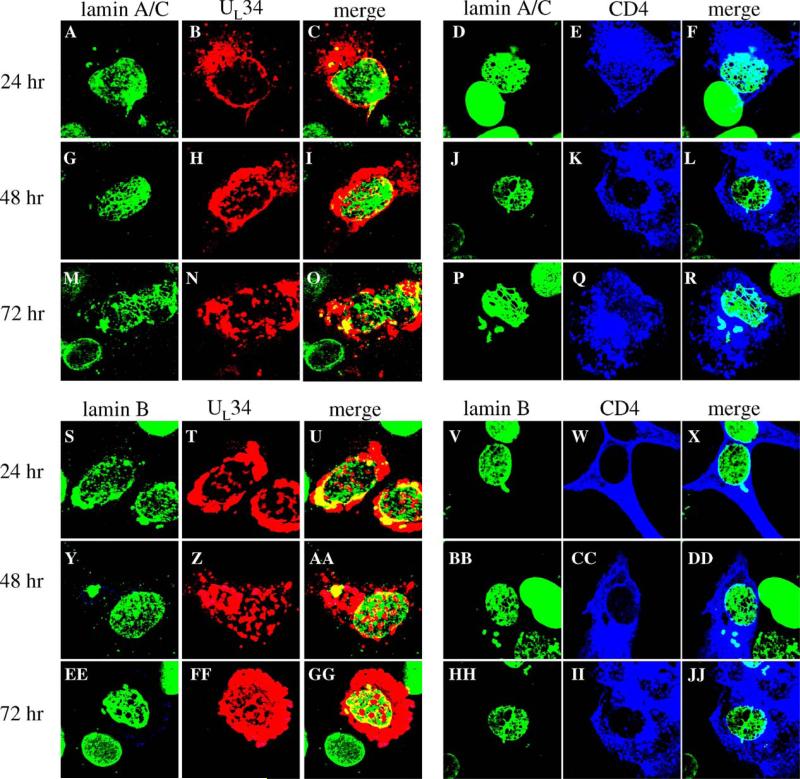
Fig. 6.
Disruption of Lamin A/C and lamin B localization by expression of transfected UL34 or CD4 genes. Shown are digital confocal images showing the localization of UL34, CD4 and lamin A/C or lamin B in Vero cells expressing either the UL34 or CD4 protein for various times. Green: lamins, red: UL34, blue: CD4. For all panels, Vero cells were transfected with plasmids expressing either UL34 or CD4 for 24, 48, or 72 h. Time after transfection is indicated to the left of each row of panels. The protein being detected is indicated above each column of panels.

