Fig. 8.
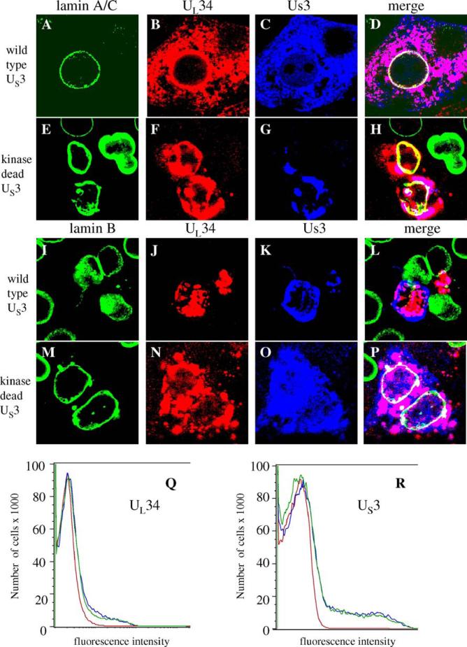
Regulation of UL34-mediated lamin disruption by co-expression of US3. For all panels, Vero cells were transfected with plasmids expressing UL34 and US3or kinase-dead US3 for 72 h. (A–Q) Digital confocal images showing the localization of UL34, US3 or kinase-dead US3 and lamin A/C or lamin B in Vero cells transfected with plasmids expressing UL34 and US3 or kinase-dead US3. All cells were transfected with UL34-expressing plasmid. The identity of the US3-expressing plasmid is indicated to the left of each row. The protein being detected is indicated above each column of panels. Green: lamins, red: UL34, blue: US3 (Q and R) Cells transfected with UL34 or US3 alone or together were analyzed by flow cytometry. (Q) UL34 staining profile for untransfected cells (red) or cells transfected with UL34 (blue) or UL34 and US3 (green). (R) US3 staining profile for untransfected cells (red) cells transfected with US3 (blue) or US3 and UL34 (green).

