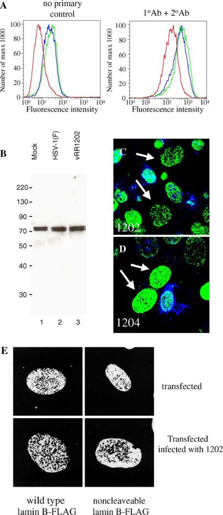
Fig. 9.
Disruptions in lamin localization are not accompanied by loss of lamin protein or cleavage of PARP. (A) Lamin A/C protein levels in Vero cells mock-infected (red), or infected with HSV-1(F) (blue) or vRR1202 (green), as analyzed by flow cytometry. The left histogram shows the results of detection with no primary antibody; the right graph shows the results of detection with both primary and secondary antibodies. (B) Lamin B protein levels in Vero cells mock-infected (lane 1), or infected with HSV-1(F) (lane 2) or vRR1202 (lane 3), as analyzed by western blot. Total cellular protein was extracted as described in Materials and methods. Coomassie staining was used to equilibrate loading (not shown). These experiments were done three times. Representative images are shown. Lane 3 is from the same blot and the same exposure as lanes 1 and 2, but was not adjacent to these lanes. (C and D) Digital confocal merged images showing the presence of cleaved PARP positive cells in Vero cells infected with vRR1202 or vR1204 at an MOI of 5 for 24 h. This experiment was done five times. A representative experiment is shown. The infecting virus is indicated on each panel. Green: lamin A/C; blue: cleaved PARP. The arrows point to cells that have perforations in the lamina, but do not stain positive for cleaved PARP. (E) Vero cells were transfected with a plasmid expressing either wild type lamin B-FLAG or non-cleavable lamin B-FLAG, then mock-infected or infected with vRR1202. The identity of the transfecting plasmid is indicated below each column. The state of infection is indicated to the right of each row.

