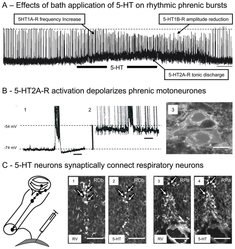
Fig. 3. 5-HT modulation of neonatal respiratory activity.

A) Traces showing in vitro phrenic bursts recorded from en bloc preparations of neonatal mice and changes evoked following bath application of 25μM 5-HT (horizontal bar). Note that 5-HT application induced complex effects characterized by an increase in phrenic burst frequency (due to activation of medullary 5-HT1AR), a tonic discharge of motoneurons (due to activation of spinal 5-HT2AR) and a reduction in phrenic burst amplitude (due to a possible presynaptic activation of spinal 5-HT1BR). Scale bar: 1 min (see Bou-Flores et al., 2000).
B) 5-HT2AR activation depolarizes phrenic motoneurons. Intracellular recording of a phrenic motoneuron before (B1) and after 5-HT application (B2). 5-HT induced a 20mV depolarization and a tonic firing of the motoneuron (time scale: 1 s; spikes truncated). B3: Immunohistochemistry reveals that neonatal phrenic motoneurons (retrogradely labelled with rhodamine) widely express 5-HT2AR (white dots). Calibration bar: 20 μm. (see also Bras et al., 2008)
C) 5-HT neurons synaptically coupled with respiratory neurons. Schematic drawing illustrates medullary neurons that project to phrenic motoneurons in neonatal mice after rabies virus (RV) inoculated in the diaphragm at P1. After RV inoculation in the diaphragm, RV progressively infected neurons controlling phrenic motoneuronal output. Thirty hours after inoculation phrenic motoneurons were labeled. After 36 hours neurons in the VRC revealed labeling and after 42 hours infected neurons were observed in histochemically identified 5-HT neurons of the raphe nuclei obscurus (1–2) and pallidus (3–4). These experiments illustrate the synaptic connectivity of raphe 5-HT neurons with phrenic premotor neurons in the VRC. Calibration bars: 100 μm. (also see Zanella et al., 2008)
