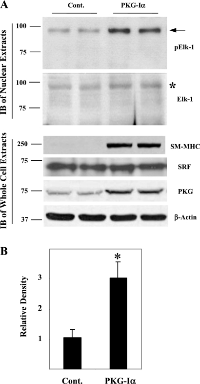
Fig. 3.
Effect of PKG-Iα expression on high molecular mass pElk-1 in rat aortic SMCs. Rat aortic SMCs at passage 3 to 5 were transfected with control pcDNA3.1 vector (Cont) or PKG-Iα-pcDNA3.1 (PKG-Iα) using Lipofectamine 2000. Stably transfected cells were selected using 500 μg/ml of G418. Cells grown to confluence were serum deprived in DMEM containing 1 mg/ml of BSA for 3 days. A: nuclear extracts or whole cell extracts were prepared and immunoblotted (IB) with indicated antibodies. Arrow on pElk-1 blot indicates high molecular mass pElk-1 *Corresponding high molecular mass Elk-1 signal on Elk-1 blot. Data are a representative of 3 experiments. SM-MHC, smooth muscle-myosin heavy chain; SRF, serum response factor. B: quantitative analysis of 3 pElk-1 blots was performed using ImageJ software. Results are expressed as means ± SD. *P < 0.01, PKG-expressing cells (PKG-Iα) vs. the control group (Cont).

