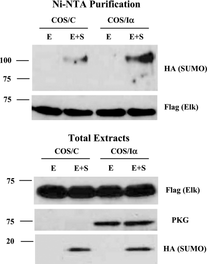
Fig. 5.
PKG-I increases Elk-1 sumoylation in vivo. His-Flag-Elk-1 (E) and hemagglutinin (HA) -SUMO-1 (S) expression vectors were transfected into control COS-7 cells (COS/C) or stably PKG-1α expressing COS-7 cells (COS/Iα). After 2 days, cells were lysed in 1 ml of 6 M guanethidine HCl, 0.1 M NaH2PO4, 0.01 M Tris·HCl (pH 8.0), and 0.3 M NaCl plus 20 mM imidazole, 20 mM β-mercaptoethanol, 15 mM N-ethylmaleimide (NEM), 5 nM calyculin A, and 1× proteinase cocktail. The lysates were mixed with 30 μl of Ni2+-nitrilotriacetic acid (Ni-NTA) agarose beads prewashed with lysis buffer and incubated overnight at 4°C. The beads were washed twice with lysis buffer and the following wash buffers: 8 M urea, 0.1 M NaH2PO4, 0.01 M Tris·HCl (pH 8.0, pH 6.3, and pH 5.9). All wash buffers contain 20 mM imidazole and 20 mM β-mercaptoethanol. After the final wash, the beads were eluted with 8 M urea, 0.1 M NaH2PO4, and 0.01 M Tris·HCl (pH 4.5) containing 200 mM imidazole and 720 mM β-mercaptoethanol. The eluates and total extracts were subjected to SDS-PAGE and immunoblotting with indicated antibodies.

