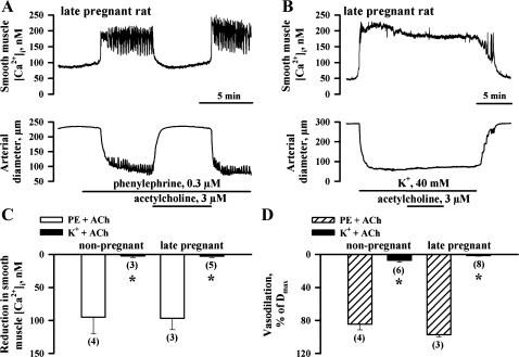
Fig. 4.
Inhibition of EDHF-mediated responses of uterine arteries by high-K+ solution. A: representative tracings showing changes in SMC [Ca2+]i and lumen diameter of the uteroplacental artery from a LP rat in response to application of 3 μM ACh. B: ACh at the same concentration failed to induce changes in SMC [Ca2+]i and the diameter of the artery preconstricted with 40 mM of K+. l-NNA (200 μM) and indomethacin (10 μM) were present throughout all experiments. C and D: bar graphs summarizing ACh-induced SMC [Ca2+]i and dilator responses of uterine arteries from NP and LP rats preconstricted with PE or high-K+ solution. Vasodilation is expressed as Dmax. Nos. in parentheses indicate the no. of tested arteries. *Significantly different at P < 0.05 (unpaired Student's t-test).

