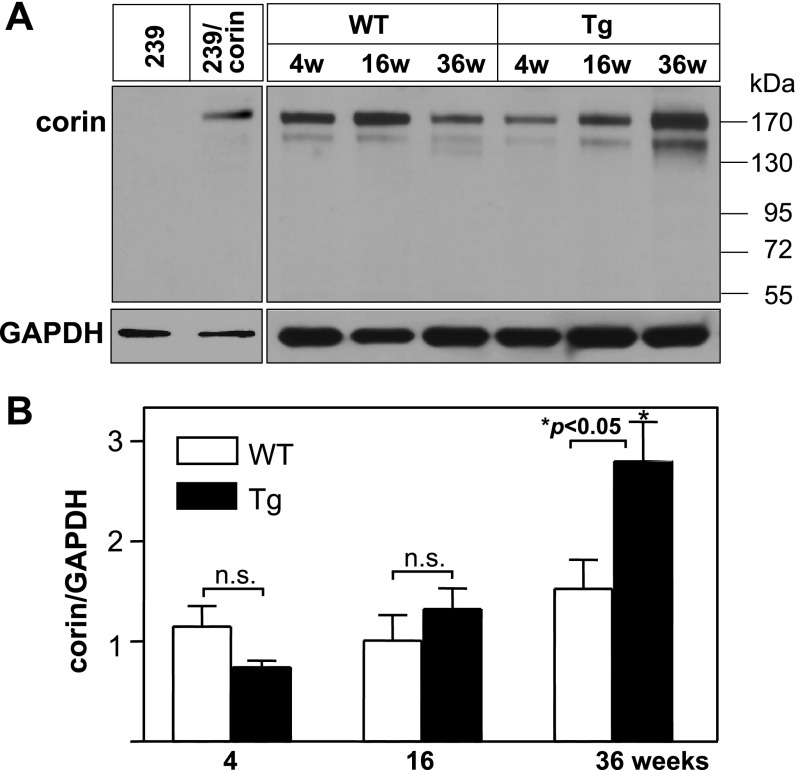
Fig. 1.
Corin protein expression in mouse hearts. A: heart membranes were from wild-type (WT) and transgenic (Tg) mice of the indicated ages (w, weeks). Corin protein was analyzed by Western blotting. Samples from parental HEK 293 cells (negative) or HEK 293 cells expressing human corin (positive) were used as controls. Blots were reprobed with an anti-GAPDH antibody, as an internal control. B: corin protein levels were quantified by densitometric analysis of Western blots. The data were from 3 independent experiments. *P < 0.05 vs. WT of the same age group; n.s., not statistically significant.

