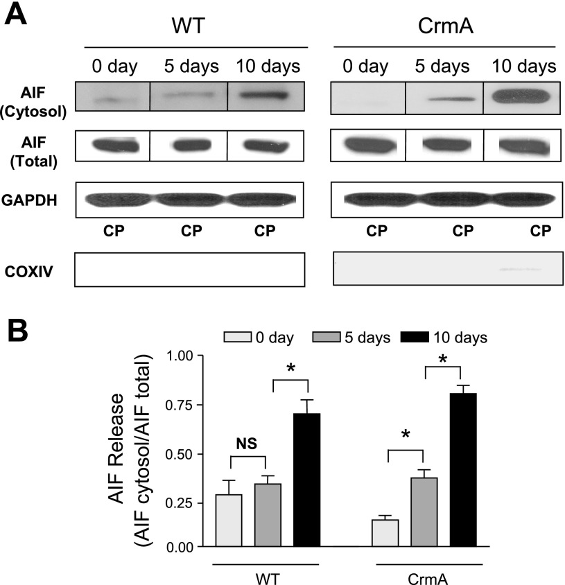
Fig. 3.
Apoptosis-inducing factor (AIF) release in Dox-induced cardiomyopathy in WT and CrmA Tg mice. A: representative Western immunoblots of AIF in the mitochondria-free cytosolic fraction and total AIF in WT and CrmA Tg mice 5 and 10 days after Dox injection. Individual representative images are identified by a line between images. The purity of the cytosolic proteins (CPs) was examined by immunoblots of (cytosolic specific) GAPDH and (mitochondrial specific) cytochrome-c oxidase subunit IV (COX IV). B: quantification of cytosolic protein content of AIF. *P < 0.05; n = 4 mice.

