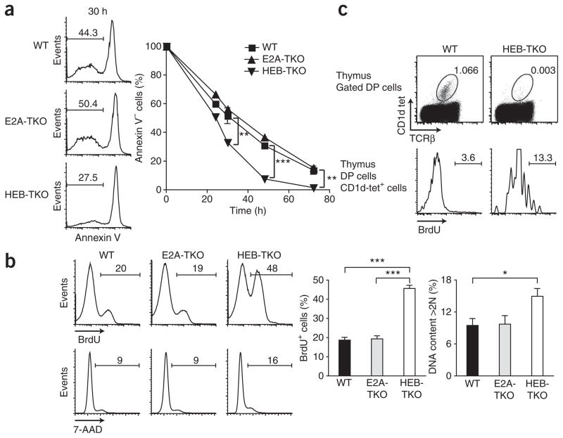
Figure 3.
HEB deficiency influences the survival and proliferation of thymocytes. (a) Annexin V expression by wild-type, E2A-TKO or HEB-TKO thymocytes cultured for 0–72 h. Numbers above bracketed lines (left) indicate annexin V–negative cells. Data are representative of five independent experiments. (b) BrdU incorporation and 7-amino-actinomycin D (7-AAD) staining of wild-type, E2A-TKO or HEB-TKO DP thymocytes after a 12-hour in vivo pulse of BrdU; numbers above bracketed lines indicate BrdU+ cells (top) or 7-AAD+ cells (middle). Data are representative of four independent experiments (average and s.e.m.). (c) BrdU incorporation by wild-type or HEB-TKO CD1d-tet+ DP thymocytes after a 3-hour pulse of BrdU. Numbers adjacent to outlined areas (top) indicate percent CD1d-tet+TCRα+ cells; numbers above bracketed lines (bottom) indicate BrdU+ cells. Data are representative of two independent experiments. *P < 0.05, **P < 0.005, ***P < 0.0005 (unpaired two-tailed t-test).

