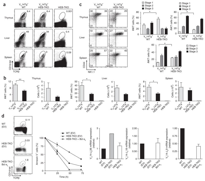
Figure 6.
Expression of a rearranged Vα14-Jα18 TCR transgene restores iNKT cell development in the absence of HEB. (a) Expression of CD1d-tet and TCRβ by Vα14tg+ wild-type, Vα14tg+ HEB-TKO and HEB-TKO lymphocytes from thymus, liver and spleen. Numbers adjacent to outlined areas indicate percent CD1d-tet+TCRβ+ cells in gate. Data are representative of five independent experiments. (b) Frequency (left) and absolute number (right) of CD1d-tet+TCRβ+ iNKT cells in thymus, liver and spleen of Vα14tg+ wild-type, Vα14tg+ HEB-TKO and HEB-TKO mice. Data are representative of two experiments (average and s.e.m.). (c) Surface expression of CD44 and NK1.1 by iNKT cells in thymus, liver and spleen of Vα14tg+ wild-type and Vα14tg+ HEB-TKO mice (left); numbers in quadrants indicate percent cells in each. Right, frequency of iNKT cells at developmental stages 1–3. *P < 0.05, **P < 0.005, ***P < 0.0005 (unpaired two-tailed t-test). Data are representative of three experiments (right, average and s.e.m. of four mice). (d) Expression of CD1d-tet and TCRβ (top left) by infected donor (CD45.2+Thy-1.1+) thymocytes in irradiated CD45.1+ hosts reconstituted with wild-type or HEB-TKO bone marrow infected with empty vector (EV; control) or HEB-TKO bone marrow infected with retrovirus encoding Bcl-xL (bottom). Right, annexin V expression by DP thymocytes obtained from the recipients described above and cultured for 0–72 h. Below, quantitative PCR analysis of transcripts of Vα14 rearrangements to Jα56, Jα18 or Jα9 segments by sorted thymocytes, presented relative to Cα transcripts. Data are representative of two experiments (top left), two independent experiments (top right) or two experiments (below; average and s.e.m. of two individual samples).

