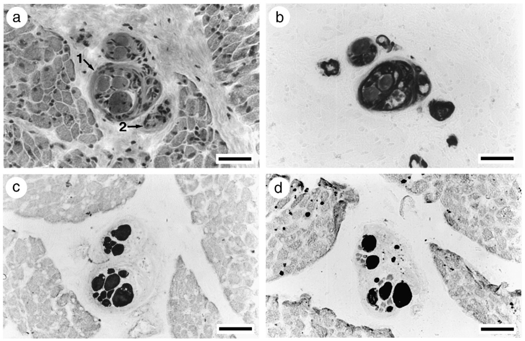
Fig. 2.
Intrafusal muscle fibers in muscle spindles from a patient in the acute phase with general myosin loss in extrafusal muscle fibers and maintained myosin expression in intrafusal muscle fibers. Note the severe muscle fiber atrophy of extrafusal compared with intrafusal muscle fibers. Serial sections stained for hematoxylin-eosin (a; arrows indicate muscle spindles), mATPase after acid preincubation (b; pH 4.6), and immunocytochemical stainings with mAbs reactive with fast (c) and slow (d) MyHCs.

