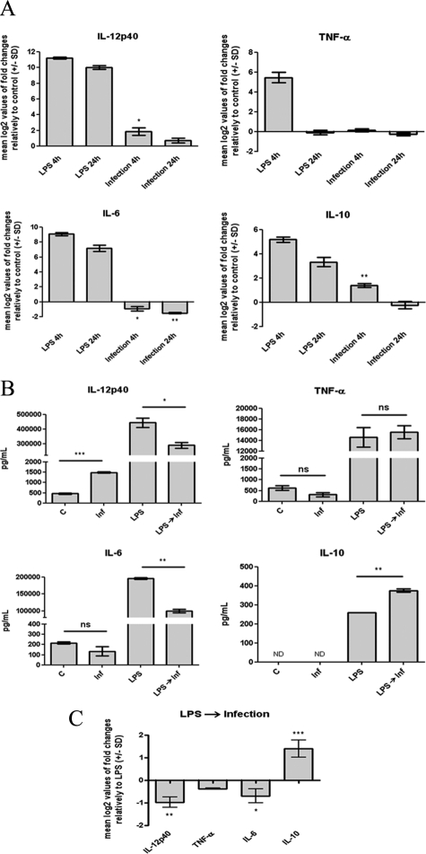
Figure 3.
Effects of L. infantum infection on BMDC cytokine production. A: Cells plated at 2 × 10⁶ cells/well were infected with L. infantum promastigotes in a 10:1 (parasites/cell) ratio or stimulated with 1 μg/ml LPS during the indicated times at 37°C with 5% CO2. The mRNA levels were assessed by real-time qPCR for IL-12p40, TNF-α, IL-6, and IL-10. Gene expression is indicated as mean log2 values of fold changes relative to untreated cells. B: The levels of IL-12p40, TNF-α, IL-6, and IL-10 were quantified by ELISA on 24-hour culture supernatants of 1 × 10⁶ BMDCs that were either infected with L. infantum promastigotes (Inf), stimulated with 1 μg/ml LPS, or pretreated for 1 hour with LPS and then infected (LPS→ Inf). C, control. C: BMDCs (2 × 10⁶/well) were stimulated with 1 μg/ml LPS or infected for 1 hour after LPS stimulation. Four hours after infection, the mRNA levels of IL-12p40, TNF-α, IL-6, and IL-10 were assessed by real-time qPCR. Gene expression is indicated as mean log2 values of fold changes relative to LPS-treated cells. Each value represents the mean ± SD from three independent experiments. *P < 0.05; **P < 0.01; ***P < 0.001; ns, not significant.

