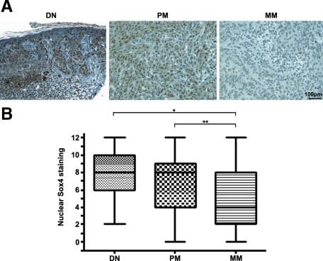
Figure 1.
Expression of Sox4 protein in cutaneous melanoma. A: Representative images of dysplastic nevi (DN) with strong nuclear staining, primary melanoma (PM) with moderate staining, and metastatic melanoma (MM) with negative staining. Scale bar = 100 μm. B: Kruskal-Wallis test for differences in Sox4 staining among DN, PM, and MM. The median is depicted as a horizontal line inside each box. *P < 0.05; **P < 0.01.

