Figure 6.
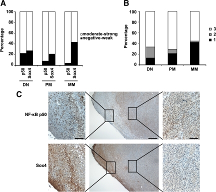
Inverse correlation between expression of Sox4 and NF-κB p50 in melanoma. A: Inverse correlation between Sox4 and NF-κB p50 expression in 169 melanocytic lesions at different stages. A significant reduced expression of Sox4 (P = 0.0260, χ2 test) and elevated expression of NF-κB p50 (P = 0.0147, χ2 test) in metastatic melanoma compared with the primary melanoma and dysplastic nevi is observed. B: Combined analysis of Sox4 and NF-κB p50 staining. Each sample was categorized based on the expression of Sox4 and NF-κB p50 as follows: 1, low (negative-weak) Sox4/high (moderate-strong) NF-κB p50; 2, high Sox4/low NF-κB p50; 3, high Sox4/high NF-κB p50. C: Representative images of serial sections of a high-risk nodular primary melanoma stained for NF-κB p50 and Sox4. Scale bar = 100 μm for left and right panels and 400 μm for the middle panels.

