Figure 4.
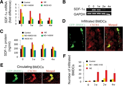
iNOS up-regulates SDF-1α expression, which, in turn, promotes BMDC infiltration. A and B show increased mRNA expression for Sdf-1α in noise-exposed mice relative to controls (n = 3; P0NE < 0.001; P1wNE < 0.001; P2wNE < 0.01; P4wNE = 0.054). C: Noise-exposed mice expressed more SDF-1α protein than unexposed controls (n = 3; P0NE < 0.01; P1wNE < 0.01; P2wNE < 0.05; P4wNE = 0.09). CXCR4 immunoreactivity was detected in migrated (D) and circulating GFP+-BMDCs (E). The merged panels in D and E show co-location of the GFP and CXCR4 labels in the GFP+- BMDCs. F: Infiltration of GFP+-BMDCs in noise-exposed and AMD3100 treated noise-exposed mice compared with controls (n = 4; P0NE = 0.46; P1wNE < 0.001; P2wNE < 0.001; P4wNE = 0.001; n = 4, P0NE+AMD3100 = 0.288, P1wNE+AMD3100 < 0.01, P2wNE+AMD3100 < 0.001, P4wNE < 0.01; C, control; NE, noise exposure).

