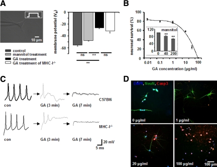
Figure 1.
A and C: GA causes a depolarization of the resting membrane potential and an immediate impairment of action potential generation in cultured hippocampal neurons. A: Resting membrane potential of cultured WT and MHC-I−deficient hippocampal neurons in the absence or presence of GA (100 μg/ml, n = 5, respectively) and its solvent mannitol (200 μg/ml, n = 3) as determined by whole-cell patch clamp recording. C: Representative recordings of action potential firing in cultured WT (upper panels) and MHC-I-deficient hippocampal neurons without GA (con), 3 and 7 minutes after incubation with GA (100 μg/ml). Black arrows indicate the time intercept between current traces. ns = not significant, **P < 0.05. B and D: GA-incubated hippocampal neurons undergo dose-dependent apoptosis within 6 hours in culture. B: GA concentration-dependent fraction of activated caspase-3+ NeuN+ hippocampal neuronal cell bodies in culture after 6 hours of incubation. (inset) Fraction of activated caspase-3+ NeuN+ hippocampal neuronal cell bodies in culture after 6 hours of incubation with the GA solvent mannitol at different concentrations (0, 40, 200 μg/ml), ns = not significant, **P < 0.05. D: Representative immunofluorescence images of cultured hippocampal neurons exposed for 6 hours to various GA concentrations. DAPI (blue), NeuN (green), activated caspase-3 (red). Scale bar = 100 μm.

