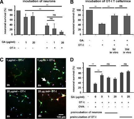
Figure 2.
Apoptosis of OVA peptide−loaded MHC-I−expressing neurons induced by OVA-specific OT-I effector T cells is reduced by pre-incubation of OT-I splenocytes but not neurons with GA. A: Fractions of activated caspase-3+ NeuN+ cell bodies of OVA peptide−loaded hippocampal neurons in culture after 6 hours of incubation with and without in vitro activated OT-I T cells in the absence or presence of two concentrations of GA (1 and 20 μg/ml). B: Fractions of activated caspase-3+ NeuN+ cell bodies of OVA peptide−loaded hippocampal neurons in culture after 6 hours of incubation with and without activated OT-I T cells either pretreated with GA (1 or 20 μg/ml) during antigen stimulation in vitro or isolated from OT-I mice pretreated with GA (100 mg/kg/d i.p.) for 30 days. ns = not significant, **P < 0.05. C: Representative immunofluorescence images of cultured hippocampal neurons incubated for 6 hours with and without in vitro activated OT-I T cells in the absence or presence of two concentrations of GA (1 and 20 μg/ml). White arrow indicates an activated caspase-3+ NeuN+ neuron. DAPI (blue), NeuN (green), activated caspase-3 (red). Scale bar = 100 μm. D: Fractions of activated caspase-3+ NeuN+ cell bodies of non-OVA peptide−loaded hippocampal neurons in culture after 6 hours of incubation with GA (1 and 20 μg/ml) alone or with OT-I T cells activated in the absence or presence of GA (1 and 20 μg/ml). ns = not significant, **P < 0.05.

