Figure 4.
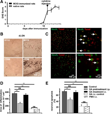
In focal EAE, lesion size and neuronal apoptosis can be limited by pretreating rats, but not by i.c. application of GA into the inflammatory lesion. A: Clinical course of MOG-induced EAE in female DA rats. B: Histological analysis of the dLGN of MOG-immunized mice, at the side of i.c. cytokine injection (right panels) and the contra-lateral control side (left panels). Stainings are shown for T cells (B115, upper panels) and macrophages (CD68, lower panels). C: Representative immunofluorescence images within the dLGN of MOG-immunized rats without (upper panel) and with GA pretreatment for 30 days (lower panel). NeuN (green), TUNEL (red), and TUNEL+ neuronal cell bodies are indicated by white arrows. Scale bar = 100 or 10 μm. D and E: Quantitative analysis of lesion size and fraction of NeuN, TUNEL+ neurons within the dLGN in MOG-immunized rats 2 days after i.c. injection of pro-inflammatory cytokines (control), in MOG-immunized rats i.p. treated with GA [2 mg/d, GA pretreatment (30 days)], in MOG-immunized rats that received i.c. injection of pro-inflammatory cytokines with GA (40 μg; GA treatment i.c.) and immunized rats that received a control injection with GA (40 μg, GA i.c. control) or PBS alone. ns = not significant, **P < 0.05.

