Figure 5.
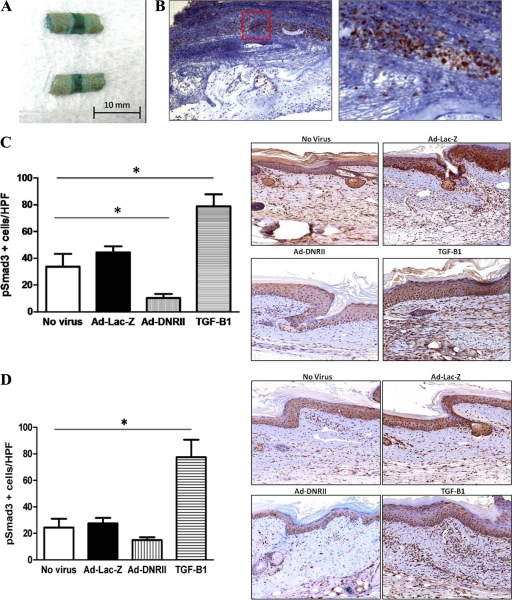
Adenoviruses can promote high-level transgene expression in mouse tail wound. A: A portion of tail transfected with Ad-Lac-Z was stained for β-galactosidase activity 1 week after surgery and transfect. The intense bluish staining, particularly at the wound, indicates increased activity and effective transfection. B: Immunohistochemistry for β-galactosidase demonstrated extensive expression in the peri-wound region 1 week after surgery and transfection with LacZ adenovirus. (Red Box: region of intense staining is magnified) C and D: Quantification of pSmad3+ cells/HPF in nonvirus, Ad-Lac-Z, Ad-DNRII, and recombinant TGF-β1 treated animals 1 (C) and 6 (D) weeks after surgery/transfection. Representative 40× images are shown to the right. Note significant decrease in the number of pSmad3+ cells/HPF in Ad-DNRII treated animals 1 week after surgery (mean ± SD; *P < 0.001). By 6 weeks, the only significant difference among groups was the number of PSmad-3 positive cells in the recombinant TGF-β1 treated group; *P < 0.001.

