Figure 6.
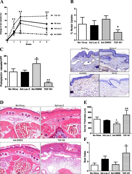
Local blockade of TGF-β1 activity decreases tail edema, increases lymphangiogenesis, and decreases tissue fibrosis. A: Tail volume measurements in nonvirus, Ad-Lac-Z, Ad-DNRII, and recombinant TGF-β1 treated animals at various time points after surgery (mean ± SD; **P < 0.01). B: Lymphoscintigraphy with Tc99 demonstrated modest, though nonsignificant, increase in nodal uptake in Ad-DNRII-treated animals. In contrasts, animals treated with recombinant TGF-β1 demonstrated markedly decreased lymphatic uptake (mean ± SEM; *P < 0.001). C: Quantification of the number of podoplanin+ lymphatic vessels in the various experimental groups 6 weeks after surgery. Mean ± SD; *P < 0.002; **P < 0.001. Representative 20× micrographs are shown to the right. D: Histological analysis of cross-sections of the tails demonstrating decreased ECM deposition and cellularity in Ad-DNRII-treated animals as compared with other groups. Representative 20× micrographs are shown. TGF-treated animals had greater swelling and lymphatic dilation compared with all other groups; 20× micrographs are presented. E: Quantitation of dermal thickness in distal tail sections 6 weeks after surgery. Treatment with Ad-DNRII decreased dermal thickness as compared with other groups. Mean ± SD; *P < 0.05. Treatment with recombinant TGF-β1 markedly increased dermal thickening; **P < 0.001. F: Quantitation of scar index by using Sirius red staining and computerized polarized light microscopy. Treatment with recombinant TGF-β1 significantly increased fibrosis; *P < 0.001. No significant differences were noted in the other groups.

