Figure 1.
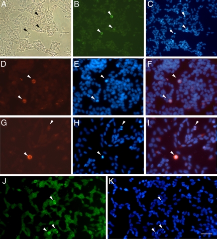
High expression of MMP-9 immunoreactivity in cells undergoing cell division. A: Phase-contrast image of SH-SY5Y neuroblastoma cells using the optical microscope. B: MMP-9 immunofluorescent staining with a mouse monoclonal antibody (Chemicon International). Arrowheads point to strongly MMP-9-immunoreactive cells (green). C: Cells undergoing mitosis as revealed with Hoechst 33258 dye (blue), which stains the dividing condensed chromosomes (arrowheads) bright blue. D–F: MMP-9 immunostaining (D, red, arrowheads) and Hoechst 33258 staining (E, blue), showing in the merged image (F) that MMP-9 immunoreactivity (red) surrounds the DNA (bright blue), in cells at metaphase (arrowheads). G–I: MMP-9 staining (G, red, arrowheads) and corresponding Hoechst 33258-stained mitotic cells (H, bright blue, arrowheads) at metaphase (bottom left) and anaphase (top right), again showing correspondence in the merged image (I). J: In situ gelatin zymography after incubation of cultured cells with fluorescein-labeled gelatin (see Materials and Methods). K: Green fluorescent staining reveals stronger gelatinase activity in cells undergoing division (arrowheads), as revealed with Hoechst 33258 dye (bright blue, arrowheads). Strong gelatinase activity is seen within mitotic cells in the area surrounding the chromosomes. Scale bars A–C = 50 μm; D–K = 25 μm.

