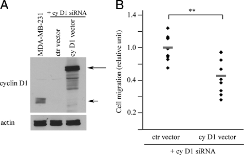
Figure 4.
Reintroduction of cyclin D1 into cyclin D1–silenced MDA-MB-231 cells. A: Western blot of MDA-MB-231 cells. First lane shows untreated cells as positive control for endogenous cyclin D1 (short arrow). Long arrow indicates reintroduced GFP-tagged cyclin D1. B: Cell migration. **P = 0.002, two-tailed Student’s t-test.

