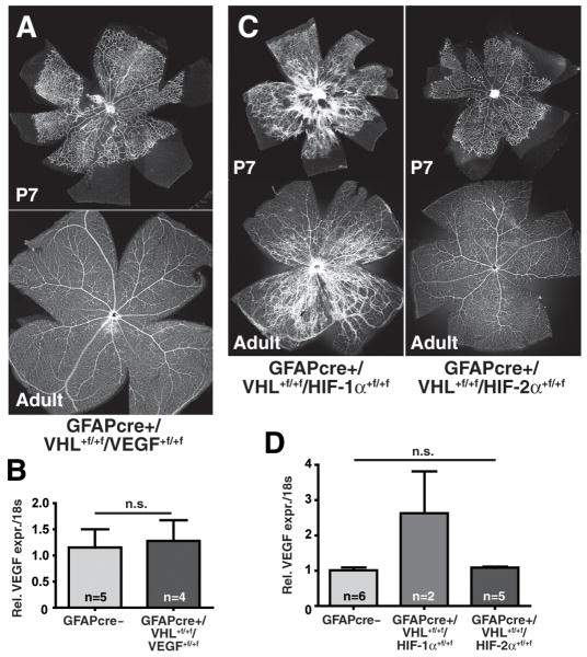
Figure 4. Pathological neovascularization in GFAPcre+/VHL+f/+f mice is driven by HIF-2α-mediated VEGF overexpression.
(A) Double knockout of VHL and VEGF in astrocytes completely rescues the hypervascular phenotype observed with Vhlh-deletion (representative picture of retina flatmounts; n=3). (B) VEGF expression levels are normal in GFAPcre+/VHL+f/+f/VEGF+f/+f mice (data represents mean ± S.E.M.). (C) Double knockout of VHL and HIF-1α in astrocytes (left) does not change the developmental hypervascularity in the retina, and the phenotype closely resembles the one of GFAPcre+/VHL+f/+f mice. In contrast, double deletion of VHL and HIF-2α (right) results in a normal development of the vasculature into adulthood (representative picture of retina flatmounts; n=3). (D) VEGF mRNA expression levels are reduced to wildtype levels by double deletion of VHL and HIF-2α, but not by VHL and HIF-1α (data represents mean ± S.E.M.).

