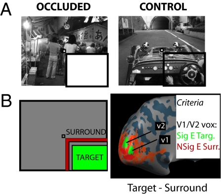
Fig. 1.
Experimental design and cortical mapping procedure. (A) The two conditions of visual stimulation used in the current experiments. In occluded trials (Left) the lower-right quadrant of the image was occluded with a uniform white field. In control trials (Right) there was no occlusion. The same three scenes were shown in both occluded and control conditions (experiments 1 and 2). The participant always had to fixate the central fixation marker (very small checkerboard) and either detect a change in frame color (experiment 1) or perform a one-back repetition task (experiment 2). Note that the black bars highlighting the occluded region are for display purposes only. The whole image spanned 22.5 × 18°, with the occluded region spanning ≈11 × 9°. (B) Cortical mapping procedure used to map the retinotopic representation of the occluded region. In a standard block-design protocol, participants were presented with contrast-reversing checkerboards in either the target region (green) or the surround region (red). We defined a patch of V1 and a patch of V2 from the contrast of target stimulus minus surround stimulus (shown here on the inflated cortical surface [left hemisphere] for one representative participant). Any vertex taken as representing the occluded portion of the main stimulus had to meet two additional constraints: a significant effect for the target stimulus alone (target > baseline; t > 1.65) and, crucially, a nonsignificant effect for the surround stimulus alone (surround stimulus = baseline; absolute t < 1.65).

