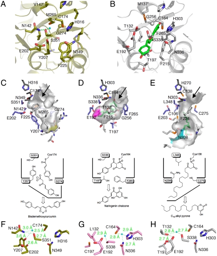
Fig. 2.
Comparison of the active-site structures of O. sativa CUS and other type III PKSs. (A and B) Comparison of the active-site structures of (A) O. sativa CUS and (B) M. sativa CHS. The naringenin molecule is shown as a green stick model. The water molecule and the hydrogen bonds are indicated with a light-blue sphere and green dotted lines, respectively. (C–E) Surface and schematic representations of (C) O. sativa CUS, (D) M. sativa CHS, and (E) M. tuberculosis PKS18. The bottom of the long pocket in CUS is indicated as a yellow surface. The bottom of the coumaroyl binding pocket and part of the side of the long-chain binding tunnel are highlighted as purple and light blue surfaces, respectively. The naringen and myristic acid bound to the active-site cavities of M. sativa CHS and M. tuberculosis PKS18, respectively, are shown as green stick models. (F–H) Close-up views of the electronic hydrogen bond networks of (F) O. sativa CUS, (G) R. palmatum BAS, and (H) P. sylvestris STS. The water molecules and the hydrogen bonds are indicated with a light-blue sphere and green dotted lines, respectively.

